

David W. McCamant
he/him/his
Associate Professor of Chemistry
PhD, University of California, Berkeley, 2004
- Office Location
- B30 Hutchison Hall
- Telephone
- (585) 276-3122
- Web Address
- Website
Research Overview
Our lab is developing new ultrafast vibrational techniques to study the nuclear geometry changes during ultrafast photochemical and photobiological reactions and the vibrational coupling that allows energy flow in small molecules. Our primary tool for these studies is a laser spectroscopy technique called femtosecond stimulated Raman spectroscopy, or FSRS, that allows the collection of high-resolution vibrational spectra with time-resolution better than 100 fs (see Figures 1 and 2 below). The power of FSRS is that it allows collection of a complete vibrational spectrum of a reacting molecule with unprecedented time resolution. This enables the determination of molecular structure as a molecule moves through transition states on the excited-state or ground-state potential energy surface. In one set of projects, FSRS is being used to determine structural changes during charge-transfer events and during the extremely short-lived electronic excited states of DNA (Figure 3). Additionally, our lab will be studying vibrational coupling by impulsively driving molecular vibrations and then observing the movement of the vibrational excitation through the molecule (Figure 4).
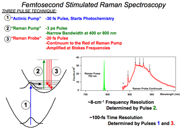
Figure 1. The laser pulses, energy diagrams and detected spectrum relevant to femtosecond stimulated Raman spectroscopy. The actinic pump is an ultrafast laser pulse in the ultraviolet or visible spectrum that initiates the photochemistry of interest. At a variable time-delay later, the Raman pump and probe obtain the Raman spectrum of the sample, which is apparent as amplification in the broad probe spectrum at Raman resonances off of the Raman pump frequency.
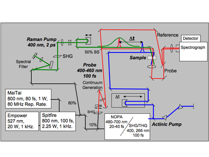
Figure 2. Schematic of the optical setup for FSRS. We start with a commercial femtosecond titanium:sapphire laser system (MaiTai, Empower, and Spitfire) and then assemble the necessary linear an nonlinear optics to produce the three necessary laser pulses.
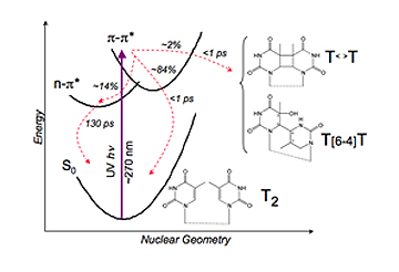
Figure 3. Electronic states involved in the photophysical relaxation of nucleic acids. Shown are the ground-state thymidine dinucleotide, T2, and the two primary photoproducts: the T<>T thymidine cyclobutane dimer, and the T[6-4]T cross-linked dimer. Conical intersections are indicated by “CI”. Adapted from Crespo-Hernandez et al., Chem. Rev. 2004, 104, 1977-2019.
Our lab is investigating the structural dynamics that occur after DNA absorbs ultraviolet light. There has been a great deal of effort by scientists around the world to determine the photophysics of this very important molecular system. Most of this work has used ultrafast electronic spectroscopy, such as transient absorption and time resolved fluorescence, to determine the kinetics of relaxation following UV photon absorption (see for instance, Crespo-Hernandez et al., Chem. Rev. 2004, 104, 1977-2019, Hare, et al., Proc. Natl. Acad. Sci. 2007, 104, 435-440 and Peon et al. Chem. Phys. Lett. 2001, 348, 255-262.) Because of these efforts, scientists now know a great deal about the kinetics of DNA photophysics; however, we still lack information on the structural dynamics, that is how the molecular geometry is distorted, during the relaxation process. Time-resolved FSRS spectra will be able to determine how the ultrafast internal conversion process is driven by changes in the excited state structure.
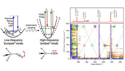
Figure 4. Schematic of 2D-FSRS experiment and the resultant 1D and 2D Raman spectra of acetonitrile.
We are developing a new multidimensional spectroscopy, entitled 2D-FSRS, in order to determine the vibrational couplings that drive energy flow through molecules. This experiment shows promise to unravel the couplings between Raman-active vibrations in much the same way that 2D-NMR techniques are used to probe magnetic couplings between nuclear spins. As shown in the experimental schematic in Figure 4, we will drive low-frequency vibrations into coherence and then probe the high-frequency Raman spectrum with a Raman pump-probe pulse pair (similar to work by Kukura et al., Phys. Rev. Lett. 2006, 96, 238303.) Anharmonic coupling between these two sets of vibrational modes will appear as peaks in a two-dimensional Raman spectrum, like the acetonitrile spectrum shown in the figure.
Research Interests
- Ultrafast spectroscopy of molecular dynamics in photochemistry
- mechanisms of vibrational relaxation
Selected Publications
- Lee, J.; Challa, J.R.; McCamant, D.W. Ultraviolet Light Makes dGMP Floppy: Femtosecond Stimulated Raman Spectroscopy of 2′-Deoxyguanosine 5′-Monophosphate. Journal of Physical Chemistry B, 121, 4722-4732, 2017. DOI: http://dx.doi.org/10.1021/acs.jpcb.7b01694.
- Ho, P.Y.; Zheng, B.; Mark, D.; Wong, W.Y.; McCamant, D.W.; Eisenberg, R. Chromophoric Dyads for the Light-Driven Generation of Hydrogen: Investigation of Factors in the Design of Multicomponent Photosensitizers for Proton Reduction, Inorganic Chemistry, 55, 8348-8358, 2016. DOI: http://dx.doi.org/10.1021/acs.inorgchem.6b00496.
- Sabatini, R.P.; Mark, M.F.; Mark, D.J.; Kryman, M.W.; Hill, J.E.; Brennessel, W.W.; Detty, M.R.; Eisenberg, R.; McCamant, D.W. A Comparative Study of the Photophysics of Phenyl, Thienyl, and Chalcogen Substituted Rhodamine Dyes. Photochemical and Photobiological Sciences,15, 1417-1432, 2016. DOI: http://dx.doi.org/10.1039/C6PP00233A.
- McCamant, D.W. Femtosecond Stimulated Raman Spectroscopy. Encyclopedia of Spectroscopy and Spectrometry, 3rd Edition, ed. George Tranter, Elsevier, 2017.
- Nganou, A.C., Kennedy, S.D., McCamant, D.W. Disagreement Between the Structure of the dTpT Thymine Pair Determined by NMR and Molecular Dynamics Simulations Using Amber 14 Force Fields. Journal of Physical Chemistry B, 120 (7), 1250-1258, 2016. DOI: http://dx.doi.org/10.1021/acs.jpcb.6b00191.
- Arpin, P., Turner, D., McClure, S.., Jumper, C., Mirkovic, T., Challa, J., Lee, J., Teng, C., Green, B., Wilk, K., Curmi, P., Hoef-Emden, K., McCamant, D.W., Scholes, G. Spectroscopic Studies of Cryptophyte Light Harvesting Proteins: Vibrations and Coherent Oscillations. Journal of Physical Chemistry B, 2015, 119 (31), 10025–10034, 2015. DOI: http://dx.doi.org/10.1021/acs.jpcb.5b04704.
- Zheng, B., Sabatini, R. P., Fu, W.-F., Eum, M.-S., Brennessel, W. W., Wang, L., McCamant, D.W. and Eisenberg, R. Light-driven generation of hydrogen: New chromophore dyads for increased activity based on Bodipy dye and Pt(diimine)(dithiolate) complexes. Proceedings of the National Academy of Sciences of the USA,112 (30), E3987–E3996, 2015. DOI: http://dx.doi.org/10.1073/pnas.1509310112.
- Dunlap, B., Richter, P., McCamant, D.W. Stimulated Raman Spectroscopy using Chirped Pulses. Journal of Raman Spectroscopy, 45 (10), 918-929, 2014. DOI: http://dx.doi.org/10.1002/jrs.4578.
- Sabatini, R. P., Eckenhoff, W. T., Orchard, A., Liwosz, K. R., Detty, M. R., Watson, D. F., McCamant, D.W., Eisenberg, R. From Seconds to Femtoseconds: Solar Hydrogen Production and Transient Absorption of Chalcogenorhodamine Dyes. Journal of the American Chemical Society, 136, 7740-7750, 2014. DOI: http://dx.doi.org/10.1021/ja503053s.
- Lee, J., Challa, J.R., McCamant, D.W. Pump Power Dependence in Resonance Femtosecond Stimulated Raman Spectroscopy. Journal of Raman Spectroscopy, 44, 1263-1272, 2013. DOI: http://dx.doi.org/10.1002/jrs.4354.
- Rhinehart, J.M., Challa, J.R., McCamant, D.W. Multimode Charge-Transfer Dynamics of 4-(Dimethylamino)Benzonitrile Probed with Ultraviolet Femtosecond Stimulated Raman Spectroscopy. Journal of Physical Chemistry B, (Part of the “Richard A. Mathies Festschrift”), 116, 10522-10534, 2012. DOI: http://dx.doi.org/10.1021/jp3020645.
- McCamant, D.W., A re-evaluation of rhodopsin’s relaxation kinetics determined from femtosecond stimulated Raman lineshapes. Journal of Physical Chemistry B, 115, 9299-9305, 2011. DOI: http://dx.doi.org/10.1021/jp2028164.
- Mehlenbacher, R., Lyons, B., Wilson, K., Du, Y., McCamant, D.W. Theoretical analysis of anharmonic coupling and cascading Raman signals observed with femtosecond stimulated Raman spectroscopy. Journal of Chemical Physics, 131, 244512, 2009. DOI: http://dx.doi.org/10.1063/1.3276684.
- Wilson, K., Lyons, B., Mehlenbacher, R., Sabatini, R., McCamant, D.W. Two dimensional femtosecond stimulated Raman spectroscopy: Observation of cascading Raman signals in acetonitrile. Journal of Chemical Physics, 131, 214502, 2009. DOI: http://dx.doi.org/10.1063/1.3263909.